Body visuializer – Body Visualizer applications are revolutionizing how we interact with and understand the human body. From detailed anatomical models to dynamic representations of physiological processes, these tools offer unprecedented clarity and insight across various fields. This exploration delves into the technology behind body visualizers, their diverse applications, and the exciting future advancements on the horizon.
We will examine the different types of body visualizers available, the technologies employed in their creation (such as 3D modeling and augmented reality), and their impact on healthcare, fitness, fashion, and gaming. A comparative analysis of various applications will highlight their strengths and target audiences, providing a comprehensive overview of this rapidly evolving technology.
Body Visualizers: A Comprehensive Overview: Body Visuializer
Body visualizers are sophisticated applications leveraging advanced technologies to create three-dimensional representations of the human body. These visualizations serve diverse purposes across various industries, from healthcare and fitness to fashion and gaming. This article delves into the core functionality, technological underpinnings, applications, and future trends of body visualizers.
Defining “Body Visualizer”
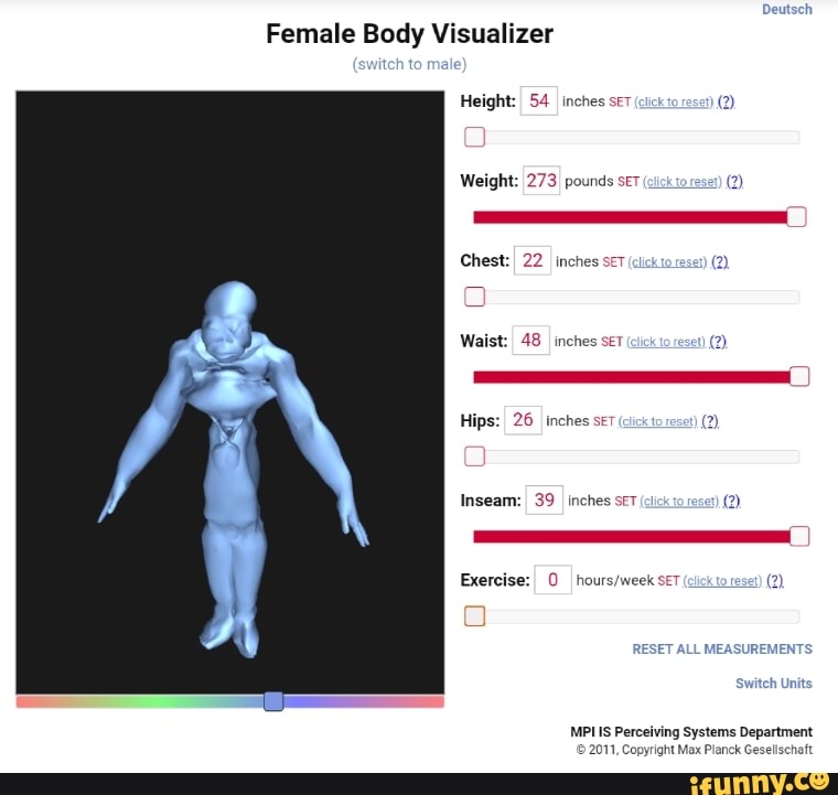
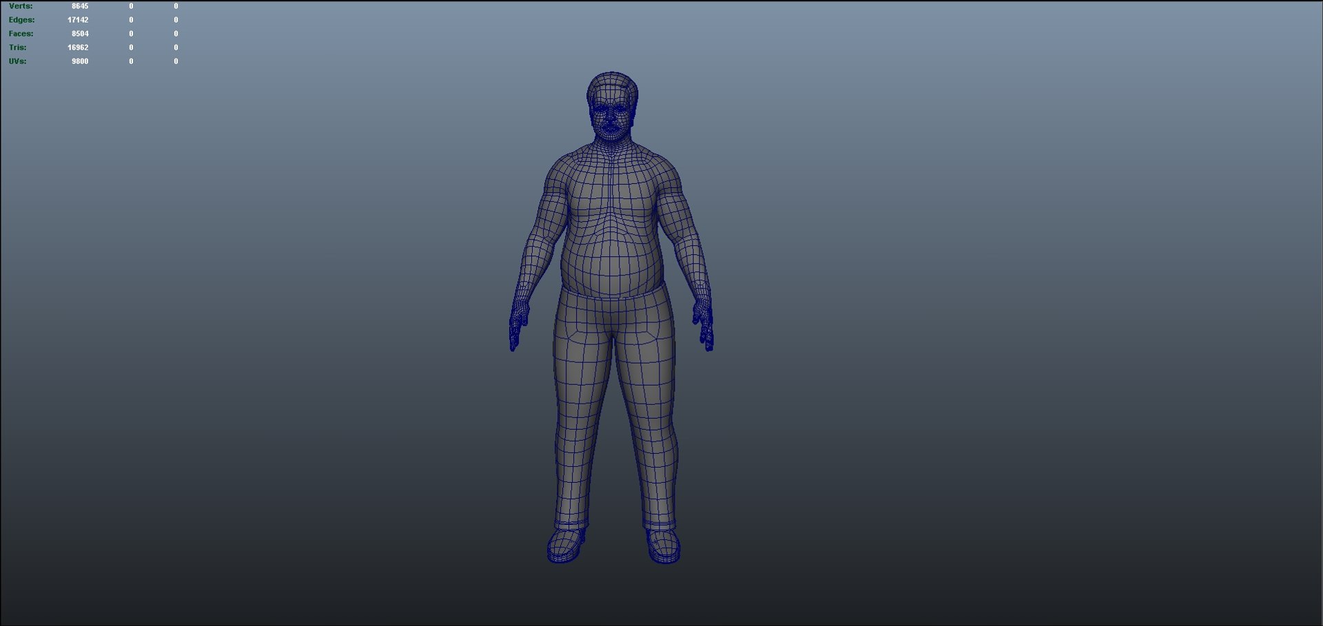
A body visualizer is a software application that generates three-dimensional (3D) models of the human body, often based on scanned data or anatomical information. The core functionality involves processing input data—which could range from medical scans to motion capture data—to create interactive and visually rich representations. These representations can be manipulated and viewed from various angles, allowing for detailed examination and analysis.
Different types of body visualizers cater to specific needs. Anatomical visualizers offer detailed representations of skeletal structures, muscles, organs, and other anatomical features. Medical imaging visualizers integrate data from techniques like MRI, CT, and X-ray to provide diagnostic visualizations. Fitness tracking visualizers often incorporate motion capture data to analyze posture, movement, and track fitness progress.
Common user interface elements include navigation tools (rotation, zoom, pan), measurement tools, layering options to display different anatomical structures, and annotation capabilities. Many incorporate customizable views, allowing users to focus on specific areas or systems. Some advanced applications even offer interactive simulations.
| Application Name | Key Features | Target User | Pricing Model |
|---|---|---|---|
| Anatomica Pro | Detailed anatomical models, interactive dissection, customizable views | Medical students, healthcare professionals | Subscription-based |
| BodyMetrics | 3D body scanning, body composition analysis, fitness tracking | Fitness enthusiasts, athletes | Freemium (with in-app purchases) |
| SurgicalSim | Pre-operative planning, surgical simulation, 3D model manipulation | Surgeons, medical researchers | License-based |
Technological Aspects of Body Visualizers, Body visuializer
The creation of realistic and functional body visualizers relies on a convergence of technologies. 3D modeling techniques are fundamental, transforming raw data into visual representations. Augmented reality (AR) overlays digital models onto the real world, enhancing the interactive experience. Image processing algorithms are crucial for cleaning, enhancing, and segmenting input data from various sources.
Different 3D modeling techniques offer distinct advantages and disadvantages. Polygon modeling, while versatile, can be computationally intensive for high-resolution models. Subdivision surface modeling provides smooth surfaces but may require more memory. Voxel-based modeling, common in medical imaging, offers precise representation of volumetric data.
Data acquisition and processing are critical. Data sources can include medical scans (MRI, CT, X-ray), 3D body scanners, motion capture systems, and even photographic images. Processing involves cleaning the data (noise reduction), segmentation (separating different anatomical structures), and rendering (creating the final visual representation).
The following flowchart Artikels the steps involved in processing body scan data for visualization:
Flowchart: Processing Body Scan Data
1. Data Acquisition (MRI, CT, etc.)
2. Data Cleaning (Noise reduction, artifact removal)
3. Segmentation (Separating tissues, organs, etc.)
4. 3D Model Reconstruction
5.
Texture Mapping (Adding realistic surface details)
6. Rendering (Creating the final visual representation)
7. Visualization (Interactive display, analysis tools)
Applications of Body Visualizers

Source: ifunny.co
Body visualizers find applications across numerous industries. In healthcare, they aid in surgical planning, diagnosis, and patient education. In fitness, they provide personalized feedback on posture, movement, and body composition. The fashion industry utilizes them for virtual try-ons and custom clothing design. The gaming industry employs them for character creation and realistic avatars.
Body visualizers offer a fascinating glimpse into the human form, allowing for detailed examination of anatomical structures. Understanding the visual representation of the body is crucial in various fields, including law enforcement, where accurate identification is paramount; for instance, consider the detailed images available through resources like mugshots wilmington nc new hanover which aid in identification. Returning to body visualizers, their applications extend far beyond forensic science, proving invaluable in medical research and education.
For example, in surgical planning, surgeons use body visualizers to create detailed 3D models of a patient’s anatomy, allowing for precise pre-operative planning and simulation of procedures. In rehabilitation, body visualizers track patient progress, provide feedback on movement, and help customize rehabilitation programs. The use of body visualizers in surgical planning emphasizes precision and minimizes invasiveness, while their use in rehabilitation focuses on personalized treatment and progress monitoring.
- Healthcare: Improved surgical planning, diagnostic analysis, patient education.
- Fitness: Personalized fitness tracking, posture analysis, body composition assessment.
- Fashion: Virtual try-ons, custom clothing design, personalized fitting.
- Gaming: Realistic character creation, immersive gaming experiences.
Future Trends and Developments
Future advancements in body visualization technology are likely to focus on increased accuracy, realism, and accessibility. The integration of artificial intelligence (AI) will likely enhance automated segmentation, analysis, and personalized feedback. Improvements in 3D scanning technologies will lead to higher-resolution models and faster data acquisition.
For instance, AI-powered analysis could automatically identify anomalies in medical scans, assisting in early disease detection. Advances in haptic technology could create more immersive and interactive experiences, allowing users to “feel” the virtual body. However, ethical considerations, particularly concerning data privacy and security, must be addressed.
Imagine a future where personalized medicine is routinely supported by highly accurate body visualizers. A patient’s unique anatomy is modeled in detail, allowing doctors to tailor treatments and interventions with unprecedented precision. This would revolutionize healthcare, improving outcomes and reducing risks.
Illustrative Examples
A detailed visualization of the human skeletal system would include the skull (frontal bone, parietal bones, temporal bones, occipital bone), the vertebral column (cervical, thoracic, lumbar, sacral, coccygeal vertebrae), the rib cage (ribs and sternum), and the appendicular skeleton (humerus, radius, ulna, femur, tibia, fibula, etc.). Each bone’s structure, articulation points, and function could be interactively explored.
A visualization of muscle groups would show the origin and insertion points of major muscles, such as the biceps brachii (originating on the scapula and inserting on the radius, flexing the elbow), the quadriceps femoris (originating on the pelvis and femur, extending the knee), and the gluteus maximus (originating on the pelvis and inserting on the femur, extending the hip).
Their primary functions and interactions would be clearly displayed.
A 3D model of the circulatory system would highlight the heart (with its chambers and valves), the aorta (the body’s largest artery), the pulmonary artery (carrying deoxygenated blood to the lungs), the vena cava (returning deoxygenated blood to the heart), and the major veins and arteries throughout the body. The flow of oxygenated and deoxygenated blood, and the functions of different vessels, could be visually demonstrated.
Final Thoughts
Body visualizers are transforming how we interact with and understand the human form. Their ability to provide clear, detailed representations has far-reaching implications across numerous industries, from revolutionizing surgical planning to personalizing fitness regimes. As technology continues to advance, we can expect even more realistic and accurate visualizations, unlocking new possibilities for education, research, and personalized healthcare.


